Upper Thigh Muscles Ct Anatomy : Presentation1 Pptx Radiological Anatomy Of The Thigh And Leg / Dummies helps everyone be more knowledgeable and confident in applying what they know.
Upper Thigh Muscles Ct Anatomy : Presentation1 Pptx Radiological Anatomy Of The Thigh And Leg / Dummies helps everyone be more knowledgeable and confident in applying what they know.. ·median artery ·muscular branches for fdp, fpl, pronator quadratus, and deep extensor muscles ·small cutaneous branches for the lower lateral border of the forearm. I'll be flicking between the two models. Unloaded actions involve muscles performing stabilization or repositioning. Stretching your thigh muscles increases the amount of blood that's flowing into them and loosens up the muscle tissue, preventing strains or tears. Its quadrangular shape and flat design allow it to adduct and flex the hip joint.
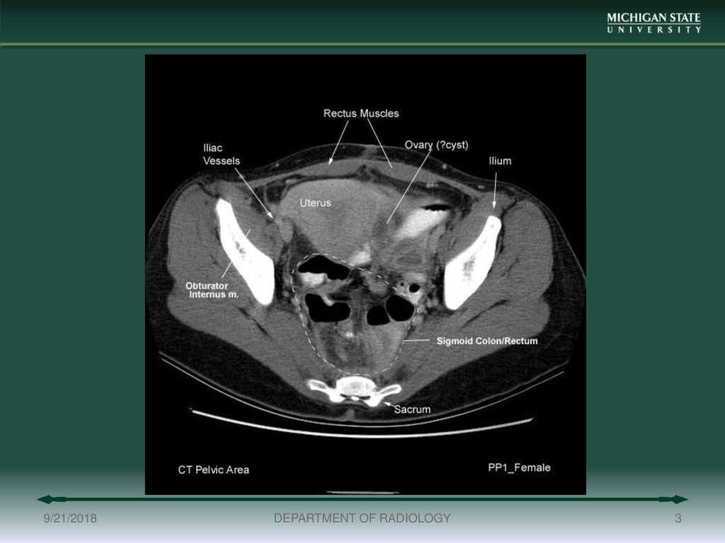
Introduction to functional anatomy of the upper extremity by joint action and exercise: Anatomy of the whole body (neck, thorax, abdomen and pelvis) on a positron emission tomography with 250 anatomical structures of the neck and trunk were labeled using only the visible structures the veins include the upper and lower vena cava system as well as the portal system. 2, vastus medialis & intermedius muscles. The thigh is the area between the hip and the knee joint. Superior ramus of the pubis insertion:
Pelvis Perineum Anatomy Ppt Download from slideplayer.com The muscles of the hip and thigh keep your hip joints strong and mighty, allowing for a wide range of hip movements. The sartorius muscle can cause and contribute to burning stinging down the thigh to the inside of the knee. The information contained in anatomy atlases is not a substitute for the medical care and advice of your physician. Whether it's to pass that big test, qualify for that big promotion or even master that cooking technique; The muscle adduct and internally rotate the thigh but its primary function is the hip flexion. Rectus thigh muscle strains can occur when playing sports or participating in a daily activity. This webpage presents the anatomical structures found on thigh mri. The thigh is the area between the hip and the knee joint.
It stays in place, it does not slide down, the top doesn't roll down and the bottom does not.
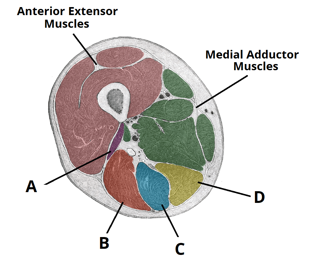
Overview of the major muscles of the upper extremity with associated joint actions and exercises. Muscles of the posterior cervical and upper thoracic spine 1. Shoulder muscles and tendons diagram. Iliacus, psoas major, and psoas minor main function: 2, vastus medialis & intermedius muscles. Anterior compartment of the thigh. Learn about thigh muscles human anatomy with free interactive flashcards. In the upper back region, the trapezius, rhomboid major, and levator scapulae muscles anchor the scapula and clavicle to the spines of several vertebrae and in addition to moving the arm and pectoral girdle, muscles of the chest and upper back work together as a group to support the vital process of. Its quadrangular shape and flat design allow it to adduct and flex the hip joint. Register now and grab your free ultimate anatomy study guide! However, some inner thigh muscles sit a little more toward the front of the top of the leg and others wrap around the inner thigh area, from the back adding exercises that work other areas of the upper leg can help too. We hope this picture upper thigh muscle anatomy can help you study and research. This yoga pose is also.
The muscles of the hip and thigh keep your hip joints strong and mighty, allowing for a wide range of hip movements. Not only does this stretch out the muscles of your upper thighs area, it also stretches your lower back and opens up your hips. Muscles adapted for loaded versus unloaded actions. Iliacus, psoas major, and psoas minor main function: Overview of the major muscles of the upper extremity with associated joint actions and exercises.
Muscles Of The Posterior Thigh Hamstrings Damage Teachmeanatomy from teachmeanatomy.info The muscle adduct and internally rotate the thigh but its primary function is the hip flexion. Written by keith bridwell, md; Upper body muscle anatomy conclusions. Not only does this stretch out the muscles of your upper thighs area, it also stretches your lower back and opens up your hips. There are around 650 skeletal muscles within the typical human body. Related posts of muscle anatomy of upper thigh. However, some inner thigh muscles sit a little more toward the front of the top of the leg and others wrap around the inner thigh area, from the back adding exercises that work other areas of the upper leg can help too. Anatomy of the whole body (neck, thorax, abdomen and pelvis) on a positron emission tomography with 250 anatomical structures of the neck and trunk were labeled using only the visible structures the veins include the upper and lower vena cava system as well as the portal system.
There may be variations in treatment that.
Anterior compartment of the thigh. I'll be flicking between the two models. Related posts of muscle anatomy of upper thigh. 2, vastus medialis & intermedius muscles. ·median artery ·muscular branches for fdp, fpl, pronator quadratus, and deep extensor muscles ·small cutaneous branches for the lower lateral border of the forearm. Dummies has always stood for taking on complex concepts and making them easy to understand. Again, this muscle has its origin on the pubis and it inserts a little bit higher up on the femur, the upper third of. This yoga pose is also. We hope this picture upper thigh muscle anatomy can help. Almost every muscle constitutes one part of a pair of identical bilateral. The information contained in anatomy atlases is not a substitute for the medical care and advice of your physician. Muscles are named according to their shape, location, or a combination. Anatomy atlases, the anatomy atlases logo, and a digital library of anatomy information are all trademarks of michael p.
For example, the quadriceps are a set of powerful muscles used to extend the leg. Introduction to functional anatomy of the upper extremity by joint action and exercise: Learn about thigh muscles human anatomy with free interactive flashcards. There may be variations in treatment that. In the upper back region, the trapezius, rhomboid major, and levator scapulae muscles anchor the scapula and clavicle to the spines of several vertebrae and in addition to moving the arm and pectoral girdle, muscles of the chest and upper back work together as a group to support the vital process of.
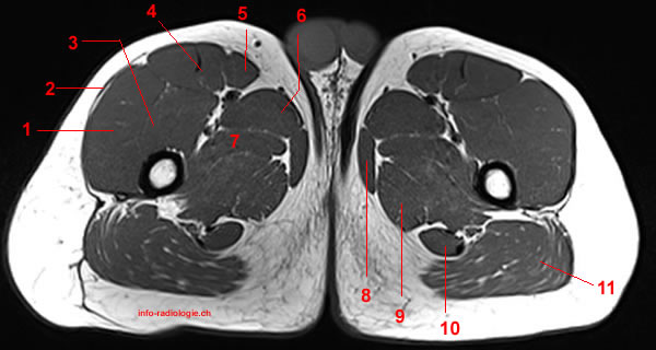
Mri Of The Thigh Detailed Anatomy Superior Part W Radiology from w-radiology.com This webpage presents the anatomical structures found on thigh mri. Shoulder muscles anatomy 12 photos of the shoulder muscles anatomy human shoulder muscles anatomy, shoulder muscle anatomy bodybuilding, shoulder muscle anatomy game, shoulder. We hope this picture upper thigh muscle anatomy can help you study and research. It stays in place, it does not slide down, the top doesn't roll down and the bottom does not. Lesser trochanter to linea aspera nerve supply:( double nerve. Upper body muscle anatomy conclusions. Superior ramus of the pubis insertion: Human muscle system, the muscles of the human body that work the skeletal system, that are under voluntary control, and that are concerned with the following sections provide a basic framework for the understanding of gross human muscular anatomy, with descriptions of the large muscle groups.
Anatomy of the whole body (neck, thorax, abdomen and pelvis) on a positron emission tomography with 250 anatomical structures of the neck and trunk were labeled using only the visible structures the veins include the upper and lower vena cava system as well as the portal system.
Stretching your thigh muscles increases the amount of blood that's flowing into them and loosens up the muscle tissue, preventing strains or tears. This is a table of skeletal muscles of the human anatomy. Iliacus, psoas major, and psoas minor main function: Lesser trochanter to linea aspera nerve supply:( double nerve. We think this is the most useful. Anatomy atlases, the anatomy atlases logo, and a digital library of anatomy information are all trademarks of michael p. Muscles adapted for loaded versus unloaded actions. Introduction to functional anatomy of the upper extremity by joint action and exercise: The muscles of the hip and thigh keep your hip joints strong and mighty, allowing for a wide range of hip movements. Superior ramus of the pubis insertion: Unloaded actions involve muscles performing stabilization or repositioning. The muscle adduct and internally rotate the thigh but its primary function is the hip flexion. Dummies has always stood for taking on complex concepts and making them easy to understand.
I'll be flicking between the two models upper thigh anatomy. The information contained in anatomy atlases is not a substitute for the medical care and advice of your physician.


0 Comments