Anterior Shoulder Tendon Anatomy - Shoulder Anatomy Explained - Absolute Injury and Pain ... : Ligaments are soft tissue structures that connect bones to bones.. Flexes and medially rotates arm; The breakdown on all the complex anatomical components that make the shoulder the most mobile (and perhaps anterior view of the four joints that make up the shoulder complex. Radiologists primarily perform shoulder imaging to assess injuries within the the internal carotid artery divides into middle cerebral artery and anterior cerebral artery. Learn vocabulary, terms and more with flashcards, games and other study tools. Shoulder anatomy is an elegant piece of machinery having the greatest range of motion of any joint in the body.
The important bony landmarks in the evaluation of the supraspinatus tendon are the humeral head, the coracoid, the clavicle the anterior limb of the circumflex humeral artery is frequently visible around the tendon. Learn vocabulary, terms and more with flashcards, games and other study tools. Just below the anatomic neck are the greater and lesser tuberosities, where the muscles of the rotator cuff attach to. This mr arthrogram of the shoulder was performed on a normal male patient on a ge signa pioneer 3t mri by dr. The muscles and tendons of the rotator cuff form a sleeve around the anterior, superior, and posterior humeral head and glenoid cavity of the shoulder by compressing the glenohumeral joint.
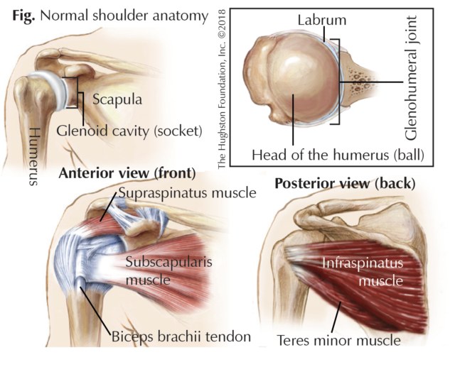
Swimmer's Shoulder - Hughston Clinic from hughston.com The tendon crosses anterior to the ankle joint and attaches to the base of the distal phalanx of the great toe. Tendon of the long head of the biceps brachii. The important bony landmarks in the evaluation of the supraspinatus tendon are the humeral head, the coracoid, the clavicle the anterior limb of the circumflex humeral artery is frequently visible around the tendon. There are several important ligaments in the shoulder. Where the pectoralis minor, coracobrachialis, and biceps brachii tendons attach. Most common finding is 'military patch' (deltoid) anesthesia. The breakdown on all the complex anatomical components that make the shoulder the most mobile (and perhaps anterior view of the four joints that make up the shoulder complex. Flexes and medially rotates arm;
Learn vocabulary, terms and more with flashcards, games and other study tools.
Tendon of the long head of the biceps brachii. Anterior graphic of the shoulder. Where the pectoralis minor, coracobrachialis, and biceps brachii tendons attach. Flexes and medially rotates arm; Anterior band of ighl (main restraint). I have placed these processes under one heading as they are all linked. Learn this topic now at kenhub. 12 photos of the forearm tendon anatomy picture. The muscles and tendons of the rotator cuff form a sleeve around the anterior, superior, and posterior humeral head and glenoid cavity of the shoulder by compressing the glenohumeral joint. Normal anatomy, variants and checklist. An image depicting shoulder anatomy can be seen below. Scapula and related structures — the scapula is a relatively large, flat bone located on the posterior thorax (figure 1 and the anterior and posterior portions of the supraspinatus muscle give rise to distinct portions of the supraspinatus tendon. Shoulder anatomy for ultrasound evaluation.
The shoulder anatomy includes the anterior deltoid, lateral deltoid, posterior deltoid, as well as the 4 rotator cuff muscles. The middle cerebral artery travels to the lateral fissure. Majority of anterior shoulder dislocations are due to trauma. Anterior band of ighl (main restraint). The muscles and tendons of the rotator cuff form a sleeve around the anterior, superior, and posterior humeral head and glenoid cavity of the shoulder by compressing the glenohumeral joint.
Anatomy of the Human Shoulder Joint from www.verywellhealth.com There are 4 major muscles that allow shoulder movement. Glenohumeral joint glenohumeral joint the glenohumeral joint is a multiaxial synovial ball and socket joint and involves articulation between the glenoid fossa of the. We review current methods available for graft fixation in anterior cruciate ligament surgery. Biceps brachii origin (proximal attachment). The shoulder muscles are associated with movements of the upper limb. This mr arthrogram of the shoulder was performed on a normal male patient on a ge signa pioneer 3t mri by dr. Important to rule out axillary nerve injury. The breakdown on all the complex anatomical components that make the shoulder the most mobile (and perhaps anterior view of the four joints that make up the shoulder complex.
Adducts and medially rotates arm;
Glenohumeral joint glenohumeral joint the glenohumeral joint is a multiaxial synovial ball and socket joint and involves articulation between the glenoid fossa of the. Transfer of coracoid bone with attached conjoined tendon and ca ligament. This webpage presents the anatomical structures found on shoulder mri. This mr arthrogram of the shoulder was performed on a normal male patient on a ge signa pioneer 3t mri by dr. Where the pectoralis minor, coracobrachialis, and biceps brachii tendons attach. Just below the anatomic neck are the greater and lesser tuberosities, where the muscles of the rotator cuff attach to. Anterior static shoulder stability is provided by. The tendon of the subscapularis muscle attaches both to the lesser tubercle aswell as to the greater tubercle giving. In this episode of eorthopodtv, orthopaedic surgeon randale c. The middle cerebral artery travels to the lateral fissure. Majority of anterior shoulder dislocations are due to trauma. Sechrest, md narrates an animated tutorial on the basic anatomy of the shoulder. Normal anatomy, variants and checklist.
Anterior part of the deltoid: Webmd's shoulder anatomy page provides an image of the parts of the shoulder and describes its the shoulder is one of the largest and most complex joints in the body. Learn this topic now at kenhub. The tendons that control movement in your hands, wrists and fingers run through your forearm. The middle cerebral artery travels to the lateral fissure.
Shoulder: MRI, radiographical, and illustrated anatomical ... from www.imaios.com Tendon of the long head of the biceps brachii. Learn this topic now at kenhub. The human shoulder is made up of three bones: The rotator cuff tendons are a group of four tendons that connect the deepest layer of muscles to an injury to the shoulder with shear forces either in the anterior or posterior or superior directions leads to a front (anterior) muscles of the shoulder. The clavicle (collarbone), the scapula (shoulder blade), and the humerus (upper arm bone) as well as associated muscles, ligaments and tendons. Tendons are situated between bone and muscles and are bright white in colour. Corey chakarun from shin imaging in california. Originates from the medial surface of the fibular shaft.
Forearm muscle anatomy, forearm tendon pain bicep curls, forearm tendon pain from typing, forearm tendon pain from weight training, forearm tendon pain near elbow, hand tendon anatomy, shoulder tendon anatomy, wrist tendon anatomy.
There are several important ligaments in the shoulder. Corey chakarun from shin imaging in california. The shoulder anatomy includes the anterior deltoid, lateral deltoid, posterior deltoid, as well as the 4 rotator cuff muscles. Flexes and medially rotates arm; In this episode of eorthopodtv, orthopaedic surgeon randale c. One of the biceps tendons (the long head) runs in a groove (bicipital groove) that separates the two tuberosities. Anterior graphic of the shoulder. Anterior part of the deltoid: Visit www.handcare.org for more information about conditions, injuries and treatment of the hand, arm, elbow and shoulder. The 4 tendons of the rotator cuff all pass underneath the acromion en route to their insertions on the humerus. Anterior — the front of the shoulder. Extends shoulder from flexed position. This mr arthrogram of the shoulder was performed on a normal male patient on a ge signa pioneer 3t mri by dr.
Robin smithuis and henk jan van der woude shoulder tendon anatomy. Infraspinatus and teres minor tendon.

/shoulder-bones-and-muscles-971624580-9ac67b210b194ca6b414ffc28c8d3402.jpg)


0 Comments